. 2023 Apr 5;17:1529. doi: 10.3332/ecancer.2023.1529

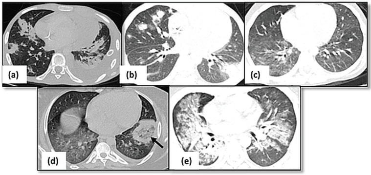
Figure 1. Pulmonary infections. (a): Axial HRCT image showing multiple consolidations in bilateral lungs with left sided pleural effusion suggestive of bacterial pneumonia. (b): Fungal infection with multiple nodules having surrounding ground glass opacity in bilateral lungs. (c): Axial HRCT image of a patient with viral pneumonia showing patchy areas of ground glass opacity in bilateral lungs. (d): Axial HRCT image showing presence of consolidation with central ground glass opacity (arrow) in left lower lobe suggestive of mucormycosis. (e): Consolidation and ground glass opacity seen in perihilar and peribronchovascular locations s/o PCP pneumonia.

Figure 1.