. 2023 Apr 5;17:1529. doi: 10.3332/ecancer.2023.1529

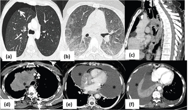
Figure 2. Pulmonary complications. (a): Axial HRCT image in a patient with osteosarcoma showing presence of subpleural metastatic nodule (arrow) in right upper lobe with right sided pneumothorax. (b): Axial HRCT image showing peripheral and lower lobe predominant ground glass opacities in bilateral lung fields in a case of Hodgkin’s lymphoma on chemotherapy s/o bleomycin toxicity. (c): Sagittal CT image of patient with esophageal cancer (asterisk) demonstrating airway invasion anteriorly (arrow). (d): Conglomerate mediastinal mass (asterisk) proven as lymphoma is seen to cause complete obliteration of SVC with multiple chest wall collaterals (arrows) – SVC syndrome. (e): Axial mediastinal window of a patient with leukemia who had massive pericardial effusion (asterisk) causing respiratory distress. (f): Axial CT iamge of lung cancer patient with gross right sided pleural effusion (asterisk) causing collpae of underlying lung. (T- trachea; E- esophagus).

Figure 2.