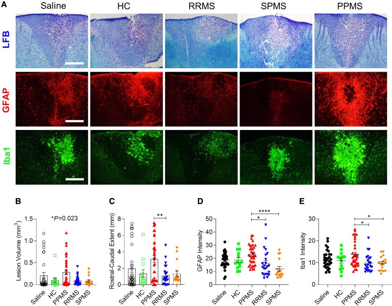
Figure 2.
PPMS CSF delays remyelination and exacerbates reactive astrogliosis and microglial activation in lysolecithin-induced lesions. (A) Representative images of the dorsal column in cervical spinal cords stained with LFB, GFAP and Iba1 at 12 days post lysolecithin injection. Mice received intrathecal injections of saline or CSF from HC (n = 3), PPMS (n = 8), RRMS (n = 7) or SPMS patients (n = 6) listed in Supplementary Table 1 at 5 days post lysolecithin injection. Saline (n = 31 mice), HC (n = 13 mice), PPMS (n = 36 mice), RRMS (n = 23 mice), SPMS (n = 16 mice). Scale bars = 100 µm. (B) Total volume of demyelination in the cervical spinal cord determined by LFB staining. (C) Rostral–caudal extent of lesion determined by LFB staining. (D) Quantification of mean fluorescence intensity of GFAP+ astrocytes throughout the lesion. (E) Quantification of mean fluorescence intensity of Iba1+ microglia throughout the lesion. Data plotted as mean ± SEM. Each point represents an individual mouse. One-way ANOVA with Bonferroni’s test (B–E). ****P < 0.0001, **P < 0.01, *P < 0.05.

