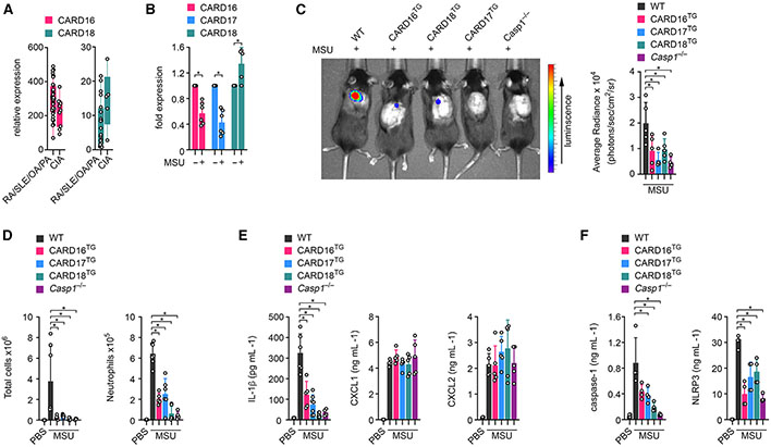
Figure 1. COPs ameliorate MSU-induced inflammation in mice.
(A) CARD16 (p = 0.0506) and CARD18 mRNA expression was determined by high-density oligonucleotide spotted microarrays (GEO: GSE36700) from synovial biopsies from patients with rheumatoid arthritis (RA), systemic lupus erythematosus (SLE), osteoarthritis (OA), and psoriatic arthritis (PA), as well as crystal-induced arthritis (CIA). n = 4–7.43,44
(B) THP-1 cells were left untreated or were treated with MSU crystals (90 μg mL−1) for 5 h, and mRNA expression of CARD16, CARD17, and CARD18 was determined by qPCR and presented as fold expression compared with untreated cells (n = 6, mean ± SD). *p < 0.05.
(C) In vivo imaging of MPO activity correlating with MSU-induced neutrophil infiltration into air pouches 7 h after MSU crystal injection (3 mg per air pouch) in wild-type (WT), CARD16TG, CARD17TG, CARD18TG, and Casp1−/− mice (left) and average radiance (right) presented as photons/s/cm2/sr (n = 4–5, mean ± SD). *p < 0.05.
(D) Total air pouch lavage cells (left) and Ly6G+ neutrophils (right) were determined by flow cytometry in mice injected with either PBS or MSU crystals (n = 4–7, mean ± SD). *p < 0.05.
(E and F) Air pouch lavage fluids were analyzed by ELISA for (E) IL-1β, CXCL1, and CXCL2 and (F) caspase-1 and NLRP3 (n = 4–7, mean ± SD). *p < 0.05. The in vivo gout model was performed twice. See also Figure S1.

