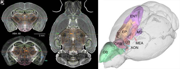
Fig. 2.
Full-brain volumetric rendering of r1CCFv3. All lines that define ROIs use the ABA color conventions. (A) The DWI in a coronal plane with ROI demarcations. (B) The FA image at the same level with four ROIs: the mediodorsal nucleus of the thalamus (MD), the caudoputamen (CP), the endopiriform nucleus (EP), and the piriform area (PIR). (C) Axial DWI of a horizontal section with corresponding borders and two ROIs labeled in common with B. The red lines in A and B and the yellow line in C define orthogonal images. (D) 3D delineations of major components of the olfactory system displayed with DSI Studio. Abbreviations—OB: olfactory bulb; AON: anterior olfactory nucleus; ENT: entorhinal area; MEA: medial amygdaloid nucleus; OT: olfactory tubule (specimen 200316-1:1)

