Fig. 4.
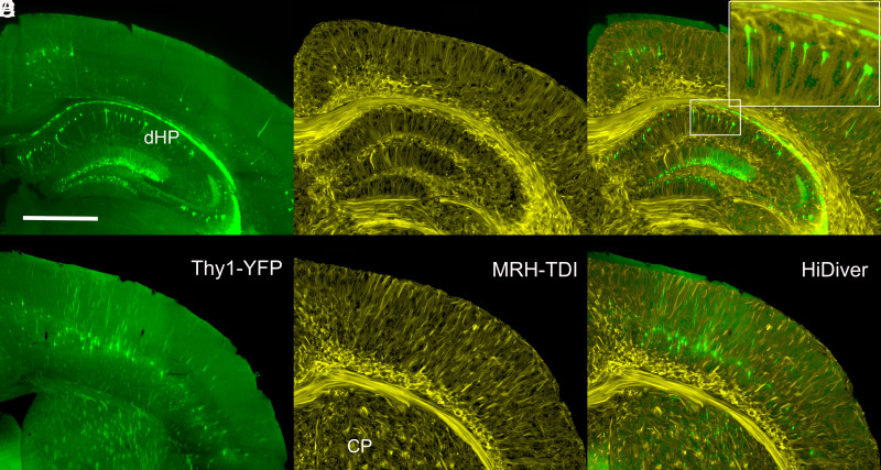
Joint LSM and superresolution TDI at two levels through cortex, dorsal hippocampus, and caudoputamen. (A) and (D) are Thy1 fluorescence images (green) from a 90-d-old B6.Cg-Tg(Thy1-YFG)HJrs/J (sample 190415-2:1). (B and E) are TDI at the same levels at a superresolution of 5 µm. (C and F) are merged HiDiver images that highlight the alignment of TDI and Thy1-positive pyramidal cells, dentate gyrus granule cells, and axon fascicles penetrating the caudoputamen. Inset in (C) is a 3× magnification of a radial section of CA1. (D–F) are LSM, TDI, and HiDiver at the level of the primary motor cortex (MOp), respectively. All images have been rendered at the same effective slice thickness of 14.4 µm. (Scale bar in A is 1 mm.) Video available @ https://bit.ly/MRHLightSheet.

