Fig. 2.
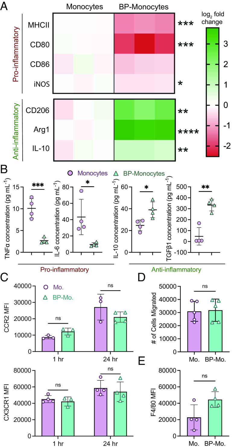
Backpacks induce anti-inflammatory phenotype in differentiating monocytes. (A) Monocytes or backpack-monocytes were cultured for 48 h and analyzed for expression of pro-inflammatory (MHCII, CD80, CD86, and iNOS) and anti-inflammatory (CD206, Arg1, IL-10) markers. Heatmap columns show data from individual replicates (n = 3), reported as log2 fold change in expression compared to the average value of the monocyte group. Raw data are in SI Appendix, Fig. S3. (B) Cytokine excretion from monocytes or backpack-monocytes after 24 h; mean ± SD (n = 3). (C) Chemokine receptor expression of monocytes (Mo.) and backpack-monocytes (BP-Mo.) at 1 h and 24 h, quantified by flow cytometry; mean ± SD (n = 3 to 4). (D) Migration was assessed using a Transwell assay, with endothelial cells seeded on 5 µm inserts, and media containing 10 ng/mL CCL2 added to the lower chamber. A total of 200k monocytes or backpack-monocytes were added into the upper chamber. The number of monocytes or backpack-monocytes in the lower chamber after 24 h was counted; mean ± SD (n = 5). (E) Monocytes or backpack-monocytes were plated and differentiated for 48 h. F4/80 expression was quantified via flow cytometry; mean ± SD (n = 4). For A, B, D, and E, data were analyzed by two-tailed Student’s t test; ns, not significant, *P < 0.05, **P < 0.01, ***P < 0.001, ****P < 0.0001. For C, data were analyzed by two-way ANOVA with Sidak’s correction; ns, not significant.

