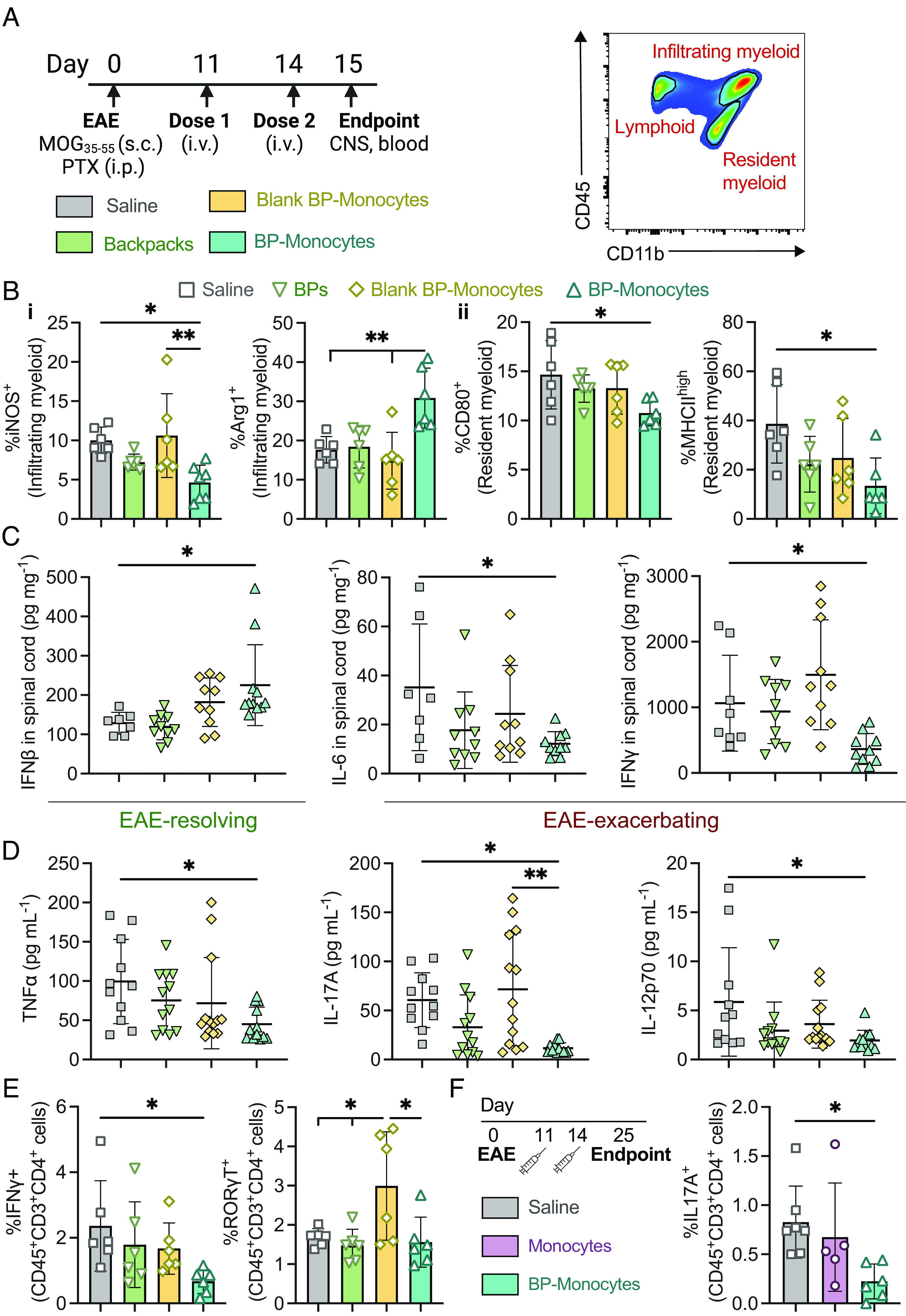
Fig. 4.

Backpack-laden monocytes modulate the CNS immune microenvironment. EAE was induced in female C57BL/6J mice. (A) The mice were treated with 3 × 10⁶ backpacks (BPs), blank backpack-monocytes, backpack-monocytes, or saline on days 11 and 14 (i.v. tail vein). On day 15, blood and CNS were harvested. Representative depiction of flow cytometry gating for distinguishing tissue-resident and tissue-infiltrating myeloid cells. (B) The spinal cord was processed into single-cell suspensions and analyzed via flow cytometry to profile the (i) infiltrating and (ii) tissue-resident myeloid cell populations; mean ± SD (n = 5). (C) Concentrations of anti-/pro-inflammatory mediators from spinal cord homogenate at day 15; mean ± SD (n = 10 to 11). (D) Serum concentrations of pro-inflammatory mediators at day 15; mean ± SD (n = 10 to 11). (E) IFNγ+ and RORγT+ CD4 T cell populations in the spinal cord at day 15; mean ± SD (n = 5). (F) EAE was induced, and mice were treated with 3 × 10⁶ monocytes or BP-monocytes or saline at days 11 and 14. At day 25, the IL-17A+ TH17 population of the blood was analyzed; mean ± SD (n = 7). For B–F, data were analyzed using one-way ANOVA with Tukey’s HSD test; ns, not significant, *P < 0.05, **P < 0.01.
