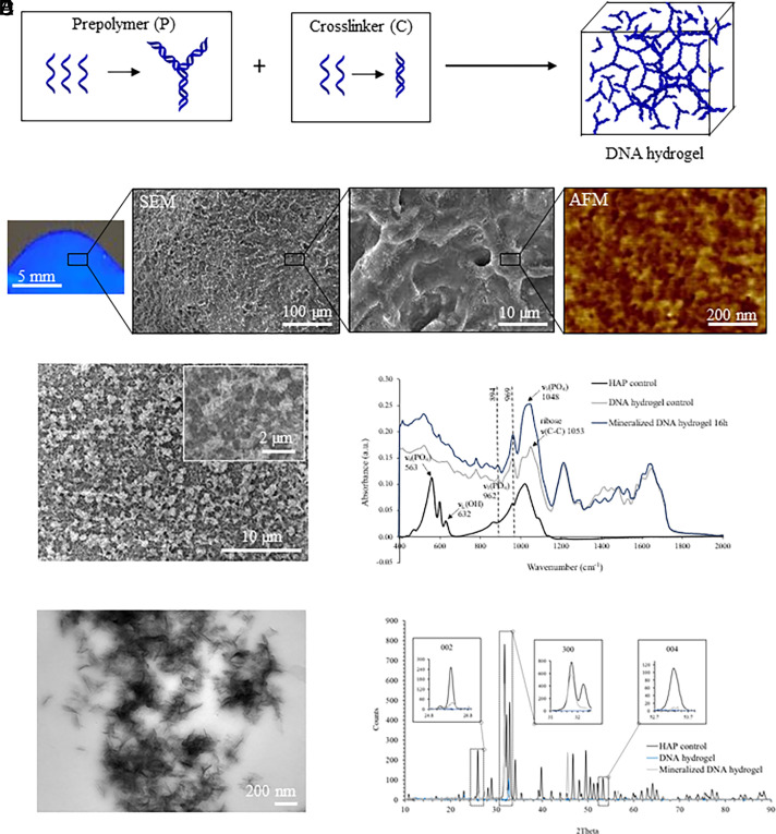
Fig. 1.
DNA hydrogel synthesis and characterization. (A) Schematic representation of DNA hydrogel formation after mixing DNA prepolymer (P) with the DNA cross-linker (C). (B) Picture of the DNA hydrogel under UV irradiation (Left), and SEM along with AFM imaging showing a fibrous porous morphology at microscale and nanoscale, respectively. (C) SEM imaging of DNA hydrogels after 16 h of mineralization. (D) Infrared spectra of mineralized DNA hydrogels confirming that the observed mineral phase is HAP. (E) TEM imaging of DNA hydrogels after 16 h of mineralization. (F) XRD spectra of DNA hydrogels after 16 h mineralization confirming that the observed mineral phase is HAP.

