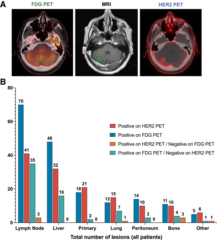
FIGURE 1.
Disease sites captured by HER2 and 18F-FDG PET. (A) 18F-FDG PET, MRI, and HER2 PET images from a patient with cerebellar metastasis. The images shown are from a patient with de novo metastatic HER2+ GEJ poorly differentiated carcinoma with mixed adeno and squamous differentiation. HER2 PET (right) demonstrated a right cerebellar metastasis (SUV 2.6) without corresponding uptake on 18F-FDG PET (left) and confirmed on brain MRI (middle). (B) Number of lesions identified by HER2 PET and 18F-FDG PET among all patients. Total number of lesions avid on HER2 PET (red) and 18F-FDG PET (blue) is shown. Total numbers of lesions better identified only on HER2 (orange) or 18F-FDG PET (green) are also shown.

