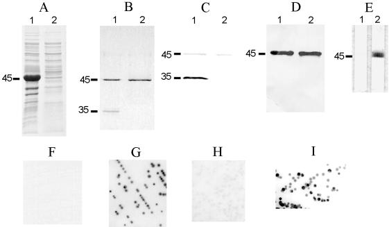
FIG. 6.
Antigenic features of rMOMP33291 and demonstration of surface-exposed conformational epitopes of MOMP. (A) Inclusion bodies (lane 1) and supernatant (lane 2) of sonicated E. coli expressing rMOMP were separated by SDS-PAGE and stained with Coomassie blue R-250. (B) rMOMP purified with Empigen BB (lane 1) or urea (lane 2) was treated in sample buffer at 25°C and then subjected to SDS-PAGE. The gel was stained with Coomassie blue R-250. (C and D) Replica samples in panel B were transferred to nitrocellulose membrane and blotted with antibodies A-8 (C) or A-5 (D). (E) rMOMP33291 purified with urea was immunostained with A-8 in the absence (lane 1) or presence (lane 2) of Empigen BB. The positions of the denatured monomer (45 kDa) and the folded monomer (35 kDa) are indicated on the left of panels A to E. (F to I) Colonies of C. jejuni strain 33291 were immunoblotted with A-5 (F), A-8 (G), A-8 absorbed with rMOMP33291 purified with Empigen (H), or A-8 absorbed with rMOMP33291 purified with urea (I).

