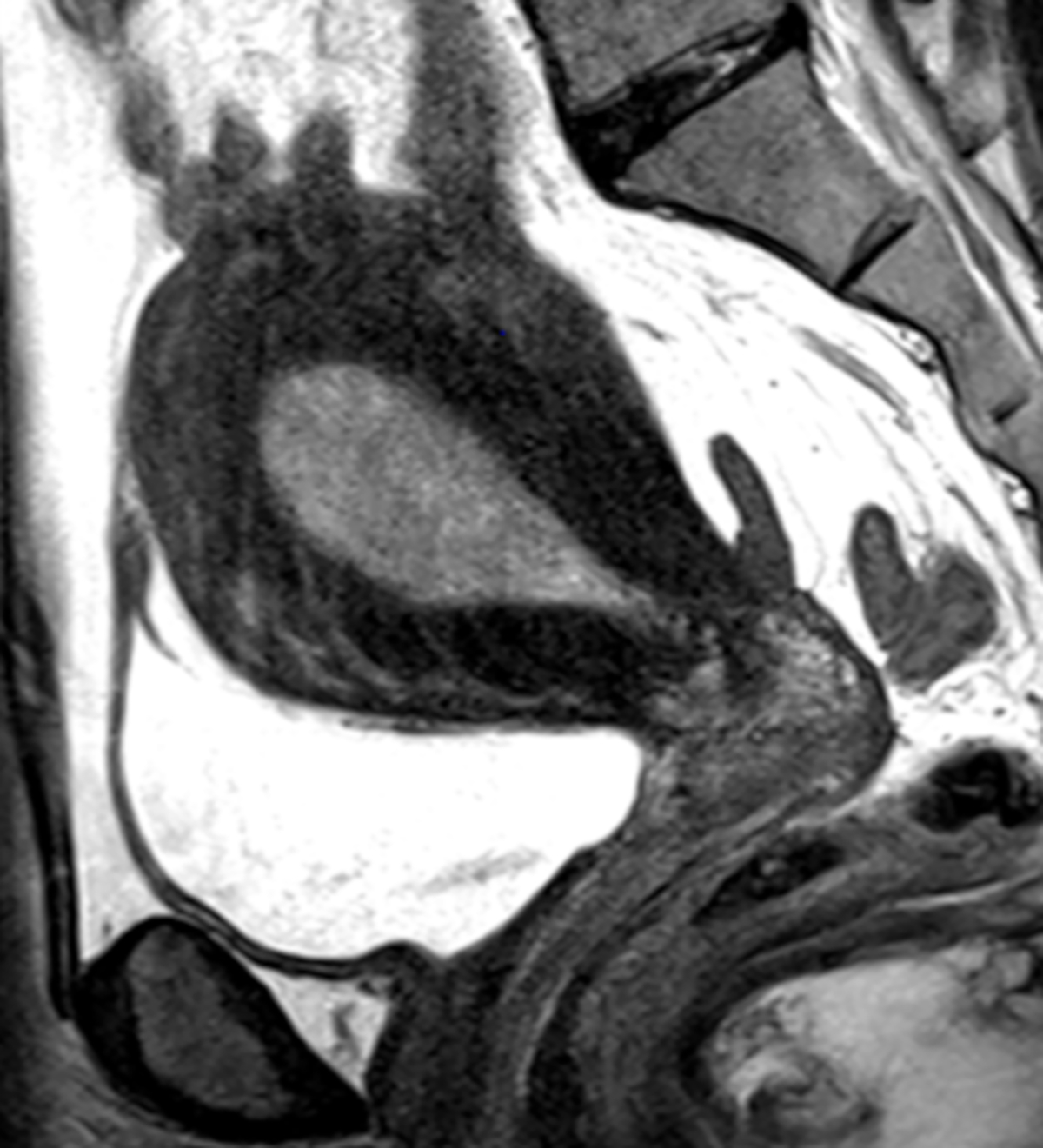
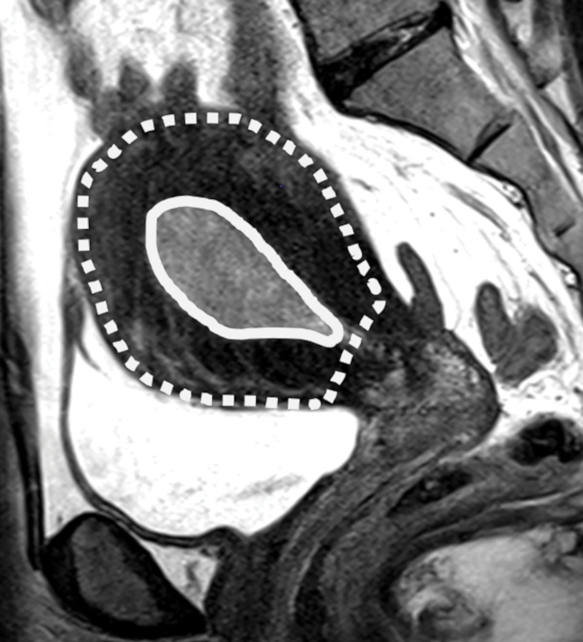
Fig. 1-.


(A) Sagittal T2-weighted image shows dilated endometrial cavity with biopsy proven endometrial cancer. (B) After manual segmentations of tumor (solid line) and the outer contour of the uterine corpus (dotted line) on each T2-weighted image, their volumes were calculated as the sum of cross-sectional volumes. Tumor volume ratio (TVR) was defined as the ratio between the tumor volume and the volume of uterine corpus. In this case, TVR was 0.11 for Reader 1 and 0.10 for Reader 2. Final surgical histology revealed superficial myometrial invasion.
