. 2023 Apr 3;59(36):5312–5328. doi: 10.1039/d3cc00573a

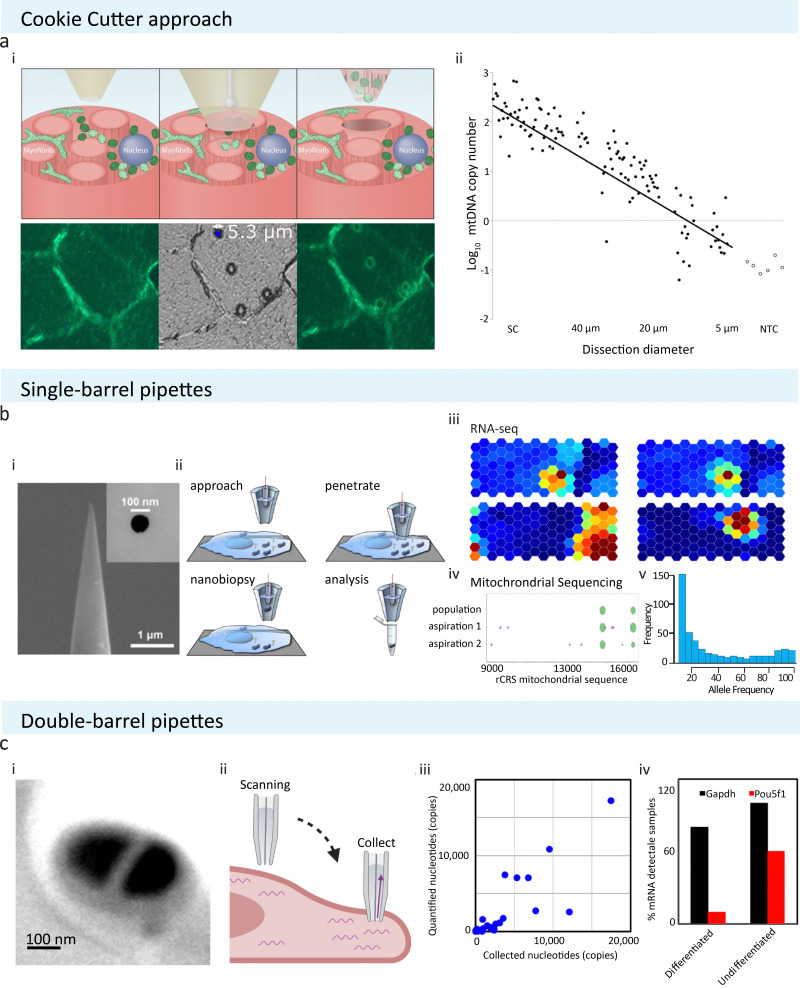
Fig. 1. Schematic of pipette-based cellular sampling technologies and downstream analysis. (a) The ‘cookie cutter’ approach from Bury et al. extracts cellular content by plunging through tissue sections. (i) Localised cellular samples are removed alongside with the micropipette after puncturing. (ii) The number of samples obtained is depends on the pore size used; with a pipette of 5 μm in size, a single mitochondrion could be extracted. Reproduced from ref. 25. Copyright 2022, Springer Nature. (b) Single-barrel pipettes from Actis et al. could be used to extract cellular samples with or without SICM. (i) SEM image of a typical single barrel with tip size of 100 nm or below are commonly utilised in extracting localised cellular samples. Reprinted (adapted) with permission from ref. 19. Copyright 2014, American Chemical Society. (ii) A schematic showing the experimental procedure of nanobiopsy when combined with SICM to obtain cellular topography. (iii) and (iv) Extracted samples have been analysed using RNA-seq, mitochondrial sequencing, or RT-qPCR to study cell-to-cell variability or single cell mitochondrial heterogeneity. Reproduced with permission from ref. 19, 28 (Copyright 2018, Elsevier), and 35 (Copyright 2017, Elsevier). (c) Double-barrel pipettes used in combination with SICM to obtain cellular contents at precise locations. (i) SEM image shows a typical dual barrel pipette applied for SICM-nanobiopsy. (ii) During SICM-single-cell biopsy, one of the two barrels of the pipette was used to acquire cell topography via SICM, and then sample extraction was done by exerting negative pressure to aspirate cellular fluid at the desired cellular location. (iii) The amount of transcripts detectable via RT-qPCR is proportional to the amount of samples collected. (iv) The dual barrel pipettes have been used to obtain subcellular amounts of samples from stem cells and study the differentiation status of stem cells by analysing differentiation marker Pou5f1 in mouse embryonic stem cells. Reprinted (adapted) from ref. 29. Copyright 2016, American Chemical Society. The schematic was created with BioRender.com.

Fig. 1