. 2023 Apr 3;59(36):5312–5328. doi: 10.1039/d3cc00573a

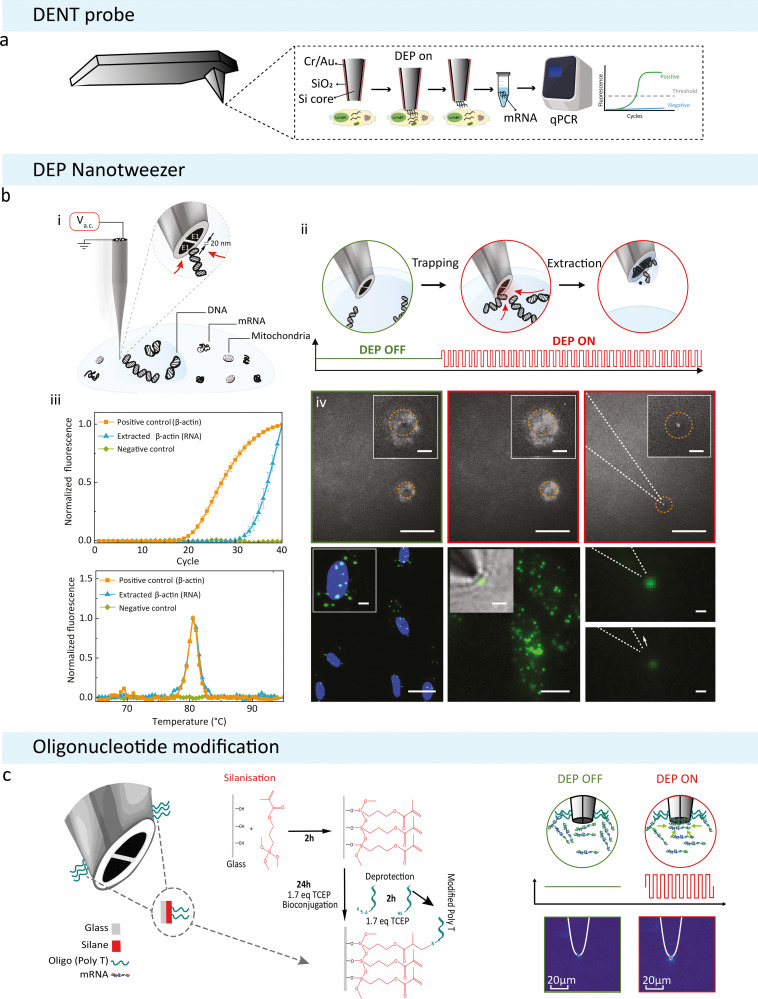
Fig. 4. DEP based subcellular sampling techniques. (a) Schematic of a modified AFM probe with DENT. Created with BioRender.com. (b) DEP Nanotweezer schematic showing how DNA can be extracted from a live cell: (i) application of an a.c. voltage on the nanotweezer generates a highly localized electric field gradient suitable for targeted molecular trapping in solution or inside a cell. (ii) Step-by-step schematics of a single cell biopsy. The tip was approached and then inserted into the cell nucleus; the application of an a.c. bias traps DNA molecules at the nanotweezer tip, and, in the final step, the nanotweezer along with the accumulated material was withdrawn from the cell. (iv) Biopsies were also performed in cells stained with a non-specific RNA dye (SYTO RNASelect). The accumulation of labelled mRNA during DEP capture results in an increase in fluorescence at the nanotweezer tip (left and middle). The mRNA can still be seen at the tip once extracted from the cell (right, scale bars = 20 μm, insets = 5 μm). (v) Targeted mRNA trapping and extraction was performed by labelling, via in situ hybridization, of individual ETS-1 mRNA molecules with fluorescein isothiocyanate (green dots) (left). A high-resolution fluorescence image of individual ETS-1 mRNA molecules (middle) along with a superimposed bright-field image is shown (inset). The application of the a.c. voltage resulted in trapping of mRNA at the nanotweezer tip (top right), which was then pulled away by the subsequent withdrawal of the nanotweezers, causing a drop in the fluorescence signal (bottom right). Scale bars: left, 25 μm (inset, 5 μm); middle, 10 μm (inset, 2 μm); right, 1 μm. Reproduced from ref. 21. (c) Updated nanotweezer system with its glass surface modified to contain an RNA Trap Poly Thymine Oligomer sequence on the tip. Modifying the surface of the glass with a broadly binding oligomer was attempted to increase the overall amount of mRNA that can be held by a single trapping event. The modification was done using click chemistry to bind the oligomer to the glass surface depicted on the left. On the right is a diagrammatic representation of the modified nanotweezers trapping free-flowing mRNA in solution. Alexa Fluor 488 labelled mRNA was extracted from MCF-7 cancer cells and diluted in 100 mM KCl. The corresponding heatmaps show the tips of the nanotweezers before and after DEP was induced.

Fig. 4