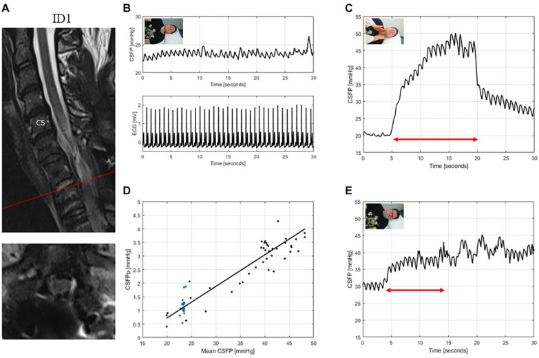
Figure 3.
T2-weighted MRI of the cervical spine for ID1 (patient with traumatic spinal cord injury, AIS: A) (A). The interpretation of the cervical MRI was challenging, but upon radiological judgement there was no clear sign of residual cord compression. CSFP and electrocardiogram (ECG) are shown during resting state (B). CSFP were normal at baseline and rise of 9.6 mmHg during Queckenstedt’s test showed a clear valvular effect (C). Cardiac-driven CSFP peak-to-valley amplitude (CSFPp) at resting state (blue dots) and during Queckenstedt’s test (black dots) is plotted against mean CSFP (D). The regression line (in black) showed a normal relative pulse pressure coefficient (RPPC-Q) equal to 0.12. Response to Valsalva maneuver is reduced with a CSFP rise of 13.0 mmHg (E).

