Figure 4.
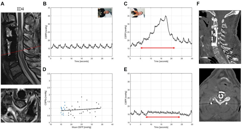
T2-weighted MRI of the cervical spine for ID4 (patient with spondylodiscitis, AIS: A) (A). This patient was tracheotomized and mildly sedated during the examination. Sagittal and axial images showed edema and were suggestive of residual cord compression related to cord swelling. CSFP was normal during resting state (B) and Queckenstedt’s test (rise 13.3 mmHg) (C). Cardiac-driven CSFP peak-to-valley amplitude (CSFPp) at resting state (blue dots) and during Queckenstedt’s test (black dots) is plotted against mean CSFP (D). The regression line (in black) was pathologically reduced with a relative pulse pressure coefficient (RPPC-Q) equal to 0.01. Response to inspiration hold maneuver is low with a CSFP rise of 1.6 mmHg (E). Sagittal and axial CT myelography reformation images of a cervical spine (F) after prolonged Trendelenburg position of 60 minutes show partial cerebrospinal fluid opacification extending to T1 (arrows), but not beyond. The constellation of findings suggests focal central C4 to C6 stenosis (asterisks), causing cranial cerebrospinal fluid flow restriction.

