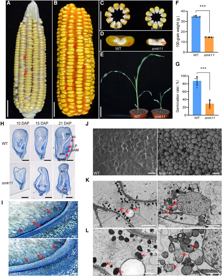
Figure 1.
Phenotypic features of maize smk11 mutant. A) A selfed ear segregates smk11 mutants at 18 DAP. Red arrows indicate the small kernels. B) A selfed ear segregates smk11 mutants at maturity. Arrows indicate the small kernels. C) Randomly selected mature smk11 and WT kernels from the segregated F2 population. D) Sections of mature WT and smk11 kernels. E) WT and smk11 mutant seedlings at 18 days after planting. F, G) Comparison of 100-grain weight (n = 8 ears) F) and germination rate (n = 5 ears) g) between WT and smk11 mutants. H) Longitudinal paraffin sections of smk11 and WT kernels at 10 DAP, 15 DAP and 21 DAP. I) The BETL cells on longitudinal paraffin sections of WT and smk11 mutant kernels at 15 DAP. Arrows indicate the BETL cells. J) Scanning electron microscopy of starch granules in the areas as boxed in Fig.1d of WT and smk11 endosperm. K) Mitochondrial ultrastructure of developing endosperms of WT and smk11 mutant at 15 DAP. Arrows indicate the Mitochondrion. L) Magnified view of mitochondria in WT and smk11 mutant. Arrows indicate the Mitochondrion. em, embryo; en, endosperm; LP, leaf primordia; RAM, root apical meristem; SAM, shoot apical meristem; sc, scutellum; BETL, BETL. Scale bars = 1 cm A-C) ; 5 mm D), 5 cm E), 1 mm H); 100 μm I); 100 μm J); 5 μm K), 2 μm L). Values are the average ± S.E. (*** P < 0.001, Student’s t test).

