Figure 4.
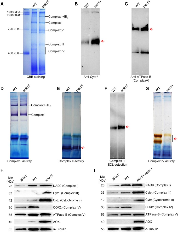
Impact on mitochondrial complexes in smk11 kernels at 15 DAP. A) BN (blue native) gel was stained with CBB. The position of mitochondrial complexes is indicated. About 100 μg of mitochondrial protein was loaded in each lane. B) BN gels transferring to PVDF membranes were probed with anti-Cytc1 (a subunit of complex III) antibody. The band of Cytc1 was indicated in arrow. C) BN gels transferring to PVDF membranes were probed with anti-ATPase-B antibody (a subunit of complex V) antibody. The band of ATPase-B was indicated in arrow. D) BN gels were used for activity staining of complex I. E) BN gels were used for activity staining of complex II. The position of mitochondrial complex II was indicated in arrow. F) BN gels transferring to PVDF membranes were detected for peroxidase activity. The position of peroxidase was indicated in arrow. G) BN gels were used for activity staining of complex IV. The position of mitochondrial complex II was indicated in arrow. H, I) Western blot analysis with antibodies against NAD9 (subunit of complex I), Cytc1 (subunit of complex III), Cytc (cytochrome c), COXII (subunit of complex IV), ATPase-B (subunit of complex V) and AOX (alternative oxidase) in mitochondrial protein from smk11 and smk11-cas9-1 kernels at 15 DAP. α-Tubulin was used as a sample loading control.

