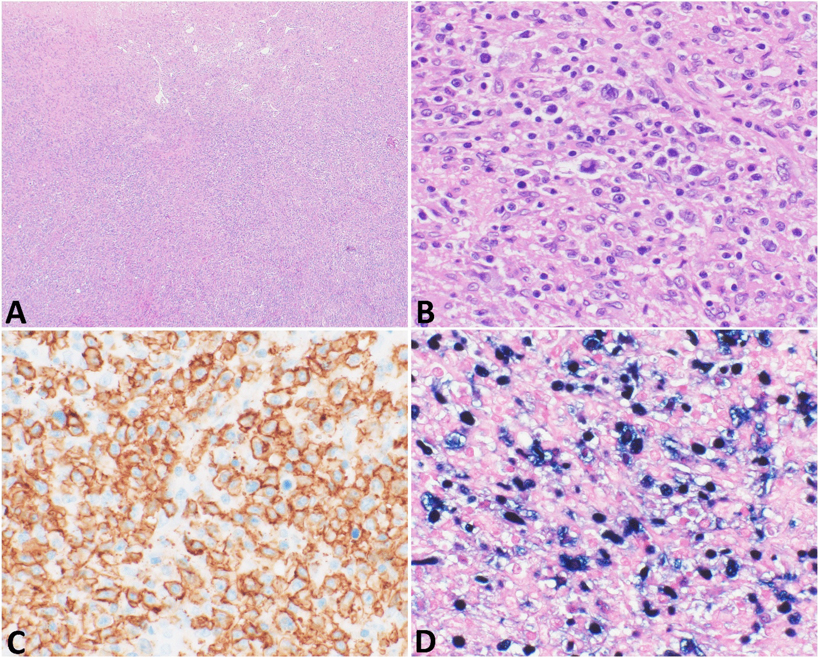
Fig. 4.

Histologic specimen. Pathological features of the liver mass. A Histologic sections show liver parenchyma is diffusely infiltrated by an atypical lymphoid population with necrosis. Magnification ×40. B The atypical lymphoid cells have large-sized nuclei, irregular nuclear contours, vesicular chromatin, distinct nucleoli, and moderate amounts of cytoplasm. Background reactive small lymphocytes and histiocytes are present. Magnification ×400. C The large, atypical lymphocytes show immunoreaction with CD20. D They are diffusely positive for EBV. Magnification ×400
