Fig. 2.
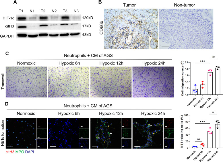
The hypoxic microenvironment of GC resulted in neutrophil recruitment and induction of NET formation. A, The expression of HIF-1α and citH3 was markedly increased in tumour tissues compared with nontumor tissues from GC patients. B, The expression of CD66b in tumour tissue and nontumor tissue samples from the same GC patient was evaluated by IHC staining. Magnification: 10 × . Scale bars: 100 μm. C, Neutrophils isolated from healthy individuals were cocultured with hypoxic-CM from GC cells, and neutrophil migration was measured by a Transwell assay. D, Representative images of NET formation by neutrophils incubated with hypoxic-CM from GC cells detected with MPO and citH3 staining. The white arrows indicate bona fide NETs. The percentage of NET releasing cells was defined as the ratio of the calculated NET releasing neutrophils to the total number of neutrophils. Magnification: 20 × . Scale bars: 50 μm. All values are the means ± SDs. ns = not significant, *p < 0.05 and ***P < 0.001

