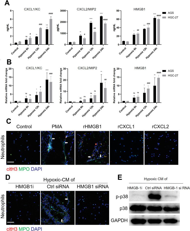
Fig. 4.
Neutrophil-attracting chemokine levels were significantly increased in GC cells incubated under hypoxic conditions. A and B, GC cells were incubated under hypoxic conditions, and the levels of neutrophil-attracting chemokines were measured by ELISA and qRT‒PCR. C, Neutrophils isolated from healthy individuals were cocultured with recombinant HMGB1, recombinant CXCL1, recombinant CXCL2 or PMA for 3 h, and NET formation was detected with MPO and citH3 staining. Images were acquired using confocal microscopy. D and E, Neutrophils were cocultured with hypoxic-CM from GC cells treated with an HMGB1 inhibitor or with hypoxic-CM from HMGB1-silenced GC cells. NET formation was evaluated by IF staining, and the level of p-p38 in neutrophils was measured by western blotting. Magnification: 20 × . Scale bars: 50 μm. All values are the means ± SDs. ns = not significant, *P < 0.05, **P < 0.01, ***P < 0.001, ****P < 0.0001 compared to control AGS cells; #P < 0.05, ##P < 0.01, ###P < 0.001, ####P < 0.0001 compared to control HGC-27 cells

