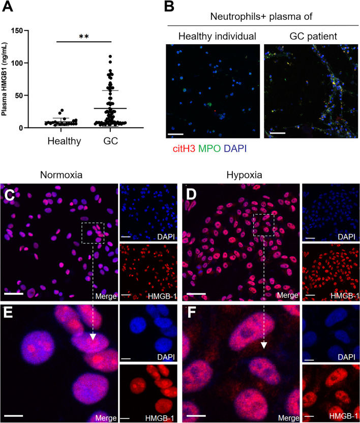
Fig. 5.
HMGB1 was translocated to the cytoplasm from the nucleus when GC cells were cultured under hypoxic conditions. A, The levels of HMGB1 in the plasma of healthy individuals (n = 20) and GC patients (n = 80) were measured by ELISA. B, Neutrophils isolated from healthy individuals were cocultured with the plasma of healthy individuals or GC patients, and NET formation was detected with MPO and citH3 staining. Magnification: 20 × . Scale bars: 50 μm. C and D, The localization of HMGB1 in GC cells under normoxia and hypoxia was determined by IF staining. Magnification: 20 × . Scale bars: 50 μm. E and F, Magnified (63x) image of HMGB1 localization in CC cells under normoxic and hypoxic conditions. Scale bars: 10 μm. All values are the means ± SDs. **P < 0.01

