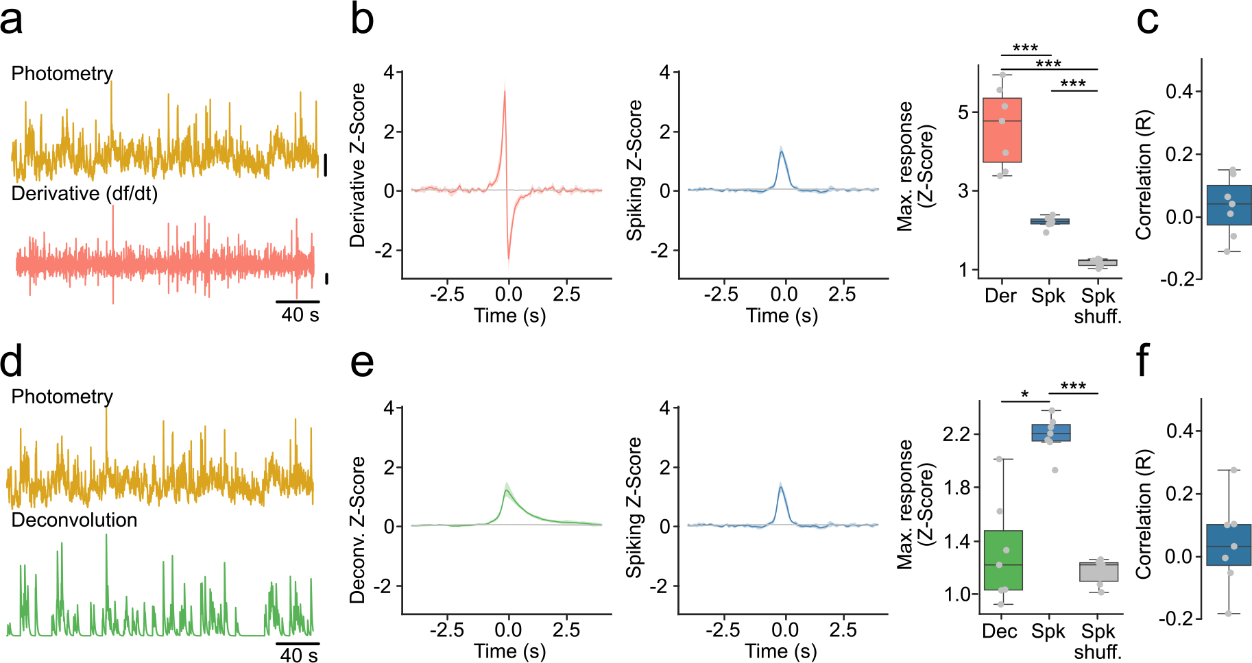
Extended Data Fig. 3 |. The time derivative and deconvolution of fiber photometry spiking activity.

(a) Example photometry trace (top) and its derivative (bottom). Vertical lines represent 2 standard deviations. (b) Derivative and spiking response around photometry transients overlapping with a spiking burst (T + B). (Left) Average derivative response. (Middle) Average population spiking response. (Right) Maximum response (n = 7 mice, F-stat = 60.80, p-value = 1 × 10⁻⁵). (c) Correlations between maximum derivative (Der) and population spiking (Spk) response around T + B. (d) Example photometry trace (top) and its respective deconvolution (bottom). Vertical lines represent 2 standard deviations. (e) Same as (b) but for deconvolution instead of derivative (n = 7 mice, F-stat = 37.74, p-value = 1 × 10⁻⁵). (f) Correlations between maximum deconvolution (Dec) and population spiking (Spk) response around T + B. Shaded regions represent 95% confidence intervals. Box plots central value denotes the median, box bounds denote upper and lower quartiles and whiskers denote ±1.5 interquartile range. For quantification of (b,e right), we used a repeated measures ANOVA with post-hoc two-tailed paired t-tests with Bonferroni corrections. * denotes p < 0.05; *** denotes p < 0.001 after corrections.
