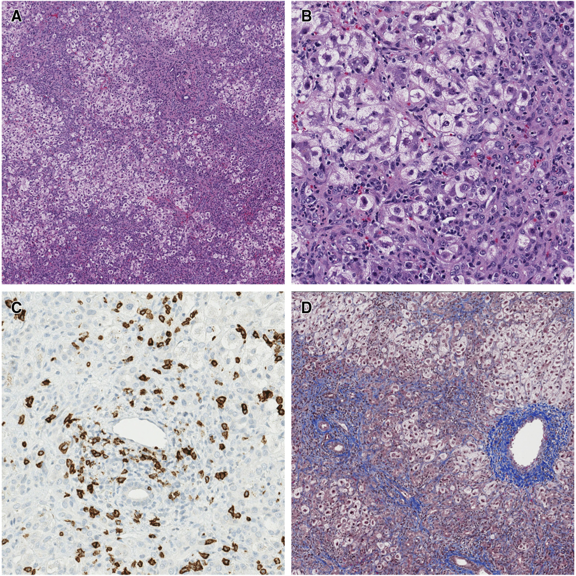
Figure 1.

Case 1 liver histology at diagnosis of liver failure. A, Extensive ballooning degeneration of hepatocytes, most prominent in zone 3 (around central veins) (stain: hematoxylin and eosin; original magnification × 40). B, Mixed portal and parenchymal inflammation including neutrophils, eosinophils, lymphocytes, and rare plasma cells. A prominent ductular reaction is also seen (stain: hematoxylin and eosin; original magnification × 200). C, Abundant CD8+ lymphocytes present within the portal tracts (CD8 immunohistochemistry-brown stain; original magnification × 200). D, Moderate amounts of portal, perisinusoidal, and central fibrosis with occasional portal-central bridging (stain: trichrome; original magnification × 40).
