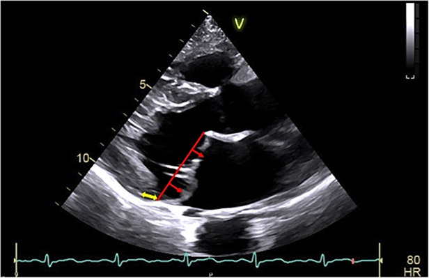
Figure 1.

Arrhythmic mitral valve prolapse by transthoracic-echocardiography. Transthoracic-echocardiographic long-axis view in end-systole with a displacement of both leaflet >2 mm (red arrow) above the plane of the annulus (red line) defining a bileaflet mitral valve prolapse with thick leaflets linked to myxomatous degeneration. Note the presence of the detachment of the posterior leaflet from the left-ventricular myocardium (mitral annular disjunction, yellow arrow) to be assessed in dynamic analysis (that is a frame by frame analysis during the entire cardiac cycle) to better ascertain the position of the posterior mitral annulus.
