
Extended Figure 2. Histopathology of hamsters infected with WT or N679K SARS-CoV-2.
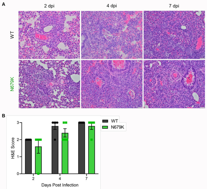
(A) H&E staining of left lung of hamsters infected with 105 pfu of WT (top) or N679K (bottom) SARS-CoV-2 at 2 (left), 4 (middle), and 7 (right) dpi. Lungs for both WT and N679K show bronchiolitis and interstitial pneumonia at 2 dpi that become more severe at 4 and 7 dpi.
(B) H&E staining of left lung of hamsters infected with 105 pfu of WT (black) or N679K (green) were scored for histopathological analysis.