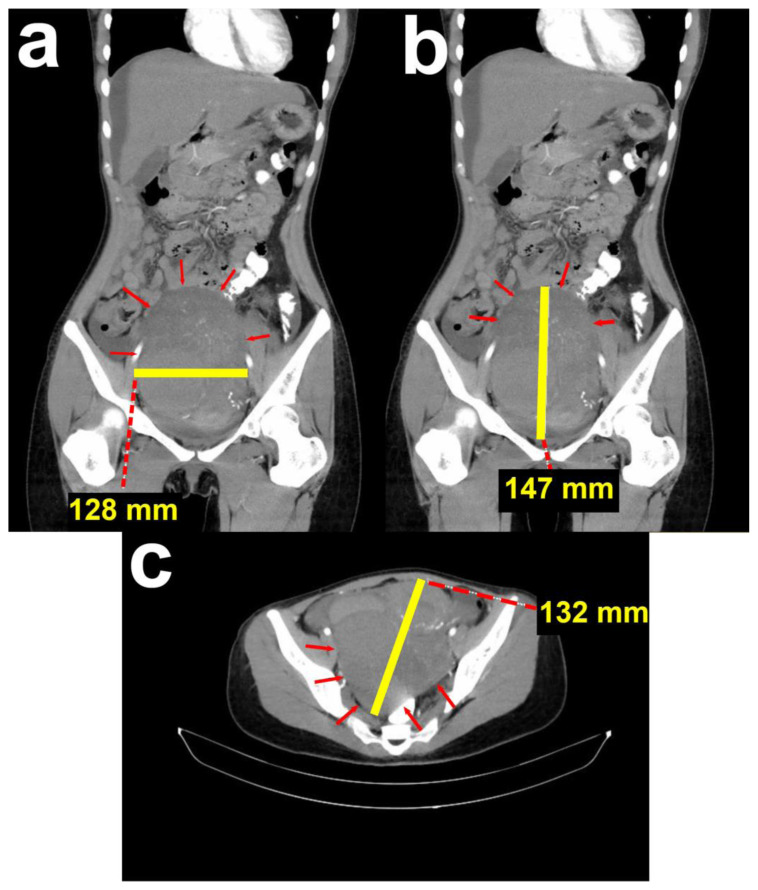
Figure 1. 24-year-old female with cystic degeneration of uterine fibroid.
Findings: CT result from the previous hospital.
(a; left; sagittal view) Mass in the pelvic cavity with a transverse diameter of 10.61 cm.
(b; middle; sagittal view) Mass in the pelvic cavity with a cranial-caudal diameter of 14.78 cm.
(c; right; axial view) Mass in the pelvic cavity with an anterior-posterior diameter of 13.28 cm.
Technique: Contrast enhanced Computed Tomography performed with acquisition of 5 mm section for all three orthogonal planes. Siemens Somatom Perspective 64 Slice scanner, 400 mAs, 120 kV, 1 mm slice thickness. Iodine contrast medium 1.5 mg/kg body weight.

