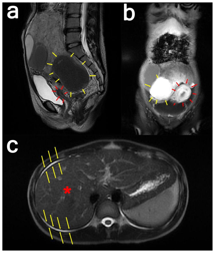
Figure 3: 24-year-old female with cystic degeneration of uterine fibroid.
Findings: (a; left) Sagittal view and (b; middle) coronal view: There is a pathological enhancement and diffusion restriction on the solid component. The cystic part shows the intensity of the blood within it. The mass appears to be pressing the uterus anteriorly.
(c; right) Axial view: The yellow arrows indicate ascites surrounding the liver.
Technique:
(a; left): Sagittal T2-weighted MRI image of the whole abdomen with contrast in a General Electric Healthcare Optima MR450w Scanner (Tesla strength = 1.5T). TR=8130.42, TE=113.40, and slice thickness = 3.0mm
(b; middle): Coronal water sequence with contrast in a General Electric Healthcare Optima MR450w Scanner (Tesla strength = 1.5T). TR=6.44, TE=3.13, and slice thickness = 8.0 mm
(c; right): Axial single-shot fast spin-echo with contrast in a General Electric Healthcare Optima MR450w Scanner (Tesla strength = 1.5T). TR=693.47, TE=92.21, and slice thickness = 8.0 mm.

