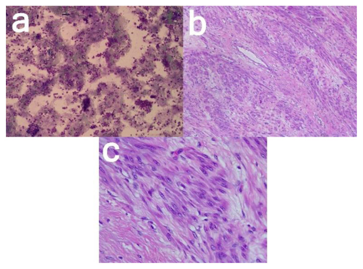
Figure 7. 24-year-old female with cystic degeneration of uterine fibroid.
Findings:
(a; left; 100× magnification) Ascites cytology
(b; middle; 100× magnification, and c; right; 400× magnification) Smooth muscle cell proliferation, round with oval nucleated, partly cigar-shaped, relatively fine chromatin.
Technique: Histopathological examination with hematoxylin and eosin stain.

