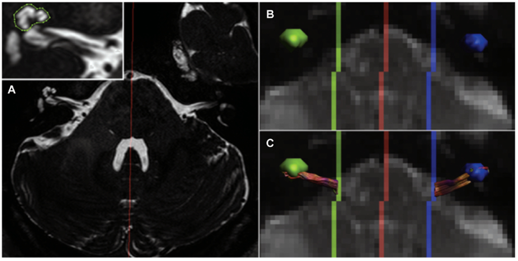
Figure 3.

Example of tractography. (A) T2-weighted structural magnetic resonance imaging used to identify the cochlear coordinates to use it as a seed point for tractography of the cochlear nerve. (B, C) Cochlear volume is transposed onto diffusion-weighted imaging, and tractography is used to connect seed points originating in the cochlea to the brainstem. Reprinted from Vos SB, Haakma W, Versnel H, et al. Diffusion tensor imaging of the auditory nerve in patients with long-term single-sided deafness. Hearing Res. 2015;323:1-8. Copyright 2015, with permission from Elsevier.
