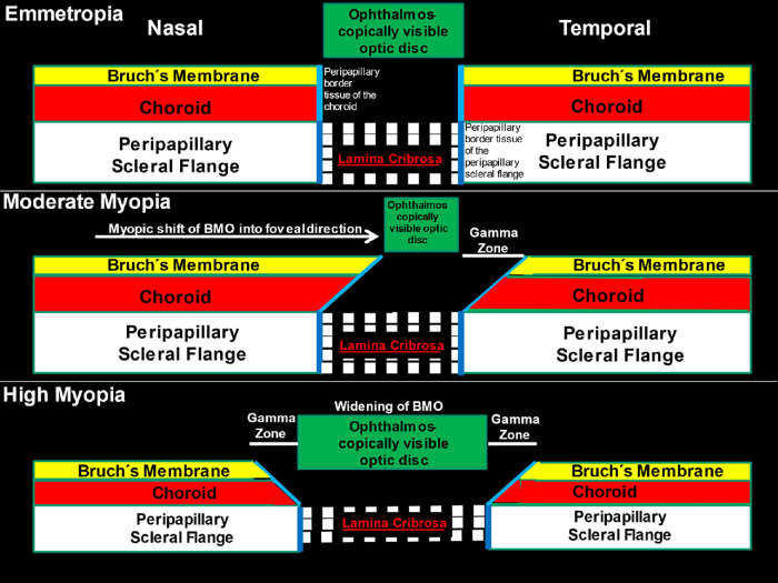
Figure 1.
Scheme showing the alignment of all three ONH canal layers (BMO, choroidal opening and peripapillary scleral flange opening, spanned by the lamina cribrosa) in emmetropic eyes (top), the shift of BMO into the temporal direction in moderately myopic eyes (middle), and the widening of BMO in highly myopic eyes (bottom). Reprinted from Jonas JB, Jonas RA, Bikbov MM, Wang YX, Panda-Jonas S. Myopia: Histology, clinical features, and potential implications for the etiology of axial elongation. Prog Retin Eye Res. 2022 Dec 28:101156. Online ahead of print. © 2022 Elsevier Ltd.

