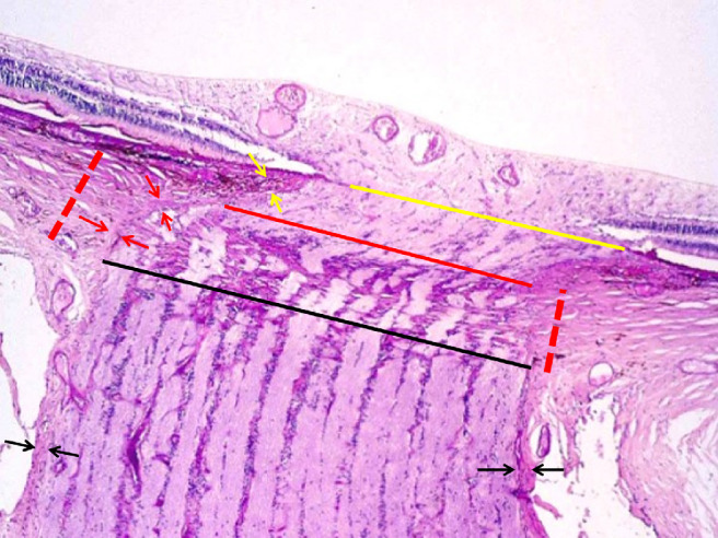
Figure 2.

Histophotograph of an ONH of a moderately myopic eye, showing the three layers of the ONH canal: the BMO (yellow line), the choroidal opening (between the yellow line and the red line), demarcated by the choroidal peripapillary border tissue (“Jacoby”) (yellow arrows), and the opening of the peripapillary scleral flange, covered by the lamina cribrosa (between the red line and the black line) and demarcated by the peripapillary border tissue of the peripapillary scleral flange (“Elschnig”) (red arrows); perforated red line: peripapillary scleral flange; black arrows: optic nerve pia mater.
