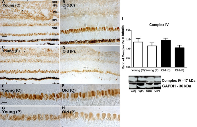
Fig 3.
A-D. Immunohistochemistry of cytochrome C oxidase subunit IV in young and old primate central (C) and peripheral (P) retina. Staining is present mostly in the ganglion cell layer (GCL), Inner plexiform layer (IPL) and outer plexiform layer (OPL, also in inner segments of the photoreceptor layer (PR). Central expression appeared heavier than that in the periphery. E-H. Images of inner segment of photoreceptors at higher magnification. Central inner segments of both age groups appear to show higher levels of expression than those in the periphery. I. Immunostaining results were confirmed with Western blot showing higher levels of complex IV in central regions. However, there were no consistent age related differences in any retinal layer. N = 5 per group. Abbreviations: GCL, ganglion cell layer. IPL, inner plexiform layer. INL, inner nuclear layer. ILM, inner limiting membrane. OPL, outer plexiform layer. ONL, outer nuclear layer. ILM, outer limiting membrane. Photoreceptors, PR. Young centre, Y(C). Young periphery Y(P). Old centre, O(C). Old periphery, O(P). See supplementary for full uncropped images.

