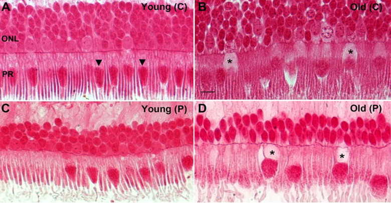
Fig 6. Representative photomicrographs showing age related changes in primate retina.
Retina from young and old primates were processed and embedded in plastic, sectioned and stained with Acid Fuchsin (mitochondrial stain). A. Photomicrograph of a young primate taken at the centre of the retina showing compact and elongated photoreceptors. The mitochondria (arrowheads) are thin, tubular and elongated. B. Photomicrograph from old primate central retina showing the photoreceptors are swollen and some of them there seem to have less Golgi and endoplasmic reticulum in the inner segment (* asterix). The mitochondria are sparse, fragmented and rounder in shape. C. Photomicrograph of young primate retina from the peripheral. The mitochondria have thin tubular and elongated shape. D. Photomicrograph of old retinae from the peripheral area. The photoreceptors are swollen and some have a void in them at the Golgi and endoplasmic reticulum area of the inner segments (* asterix). The mitochondria appear fragmented and rounder in shape.

