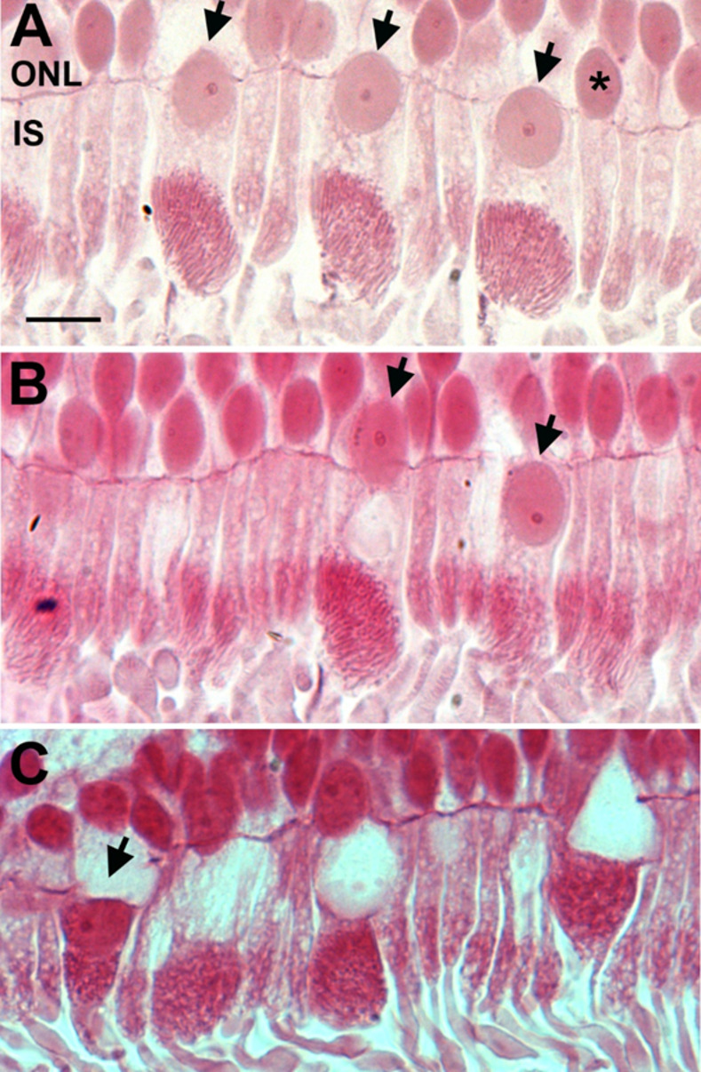
Fig 7. Displacement of nuclei from the outer nuclear layer to the inner segment.

Representative photomicrographs of old primate peripheral retina showing displacement of the nuclei from the outer nuclear layer (ONL) to the inner segment (IS) of the photoreceptors (black arrows). The retinae were sectioned at 2.5 μm and stained with Acid Fuchsin. A. Nuclei displacements seem to occur mainly in cones (black arrows) and rarely in rods (*asterix). It is observed that nuclei displacement occurs more in the old primate periphery than in young peripheral retina. B. Micrograph shows that the nucleus has completely crossed the outer limiting membrane into the inner segment, in the area of endoplasmic reticulum and Golgi (the black arrow on the right). C shows that the nuclei have migrated and is sitting on top of the mitochondria (black arrow). Scale bar = 10μm.
