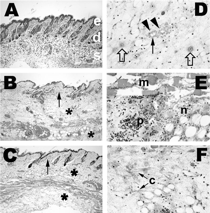
FIG. 2.
Histopathology of infection with virulent clinical and attenuated environmental V. vulnificus strains. Mice were injected i.p. with iron dextran immediately before s.c. inoculation with 102 CFU of virulent clinical strains or 105 CFU of attenuated environmental strains. Tissues were collected from each mouse 14 to 20 h after inoculation, fixed in buffered formalin, embedded in paraffin, and cut into 5-μm sections. Sections were stained with hematoxylin and eosin. Magnifications, ×100 (A through C) and ×400 (D through F). (A) Normal mouse skin. Epidermis (e), dermis with hair follicles and sebaceous glands (d), and subcutis with adipocytes, blood vessels, and muscle layer (s) are shown. (B) Skin of mouse inoculated with 500 CFU of virulent clinical strain LL728. Shown is severe edema of the subcutis (∗), extending into dermis (arrow). (C) Skin of mouse inoculated with 105 CFU of attenuated environmental strain MLT403. Shown are edema and inflammatory cell accumulation in subcutis (∗), extending into dermis (arrow). (D) Subcutis of mouse inoculated with 500 CFU of virulent clinical strain 2400112. Shown is severe edema with numerous bacteria. Degenerated small vessels with adjacent necrotic neutrophils are indicated by arrowheads, and fibrin deposition or thrombosis is indicated by a closed arrow. Cellular debris and fibrin probably representing necrotic capillaries are indicated by open arrows. (E) Subcutis of mouse inoculated with 105 CFU of attenuated environmental strain MLT367. m, necrotic s.c. muscle; p, edematous subcutis with moderate numbers of neutrophils. Many neutrophils are necrotic (n). (F) Subcutis of mouse inoculated with 500 CFU of virulent clinical strain LL728. Shown are s.c. edema with numerous bacteria as in panel D. Severity of edema is indicated by wide separation of collagen fibers (c).

