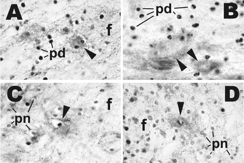
FIG. 3.
Effects of iron dextran on histopathology of V. vulnificus infection. Mice receiving iron dextran before infection were treated exactly as described in the legend to Fig. 2. Non-iron-dextran-treated mice were inoculated with 107 CFU of clinical strain LL728 or 108 CFU of environmental strain MLT403. Tissues were prepared as described for Fig. 2 and stained with hematoxylin and eosin. Magnification, ×1,000 (all panels). (A and B) Subcutis of mice inoculated with clinical strain LL728 with iron (A) and without iron (B). (C and D) Subcutis of mice inoculated with environmental strain MLT403 with iron (C) and without iron (D). There is edema with deposition of fibrin fibrils (f), degenerated but identifiable neutrophils (pd), and necrotic neutrophils represented by amorphous remnants (pn). In panels A, C, and D, there are necrotic capillaries accompanied by fibrin deposits (arrowheads). In panel B, the structures identified by arrowheads probably represent necrotic small vessels.

