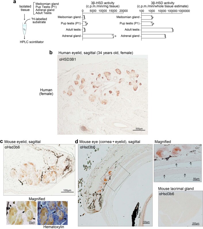
Extended Data Fig. 1. Meibomian gland 3β-HSD activity and protein localization.
a, Relative intensity of local 3β-HSD activity of pup testis, adult meibomian gland, testis, and adrenal gland, related to Fig. 1f. Tissues were isolated from 3-month-old male mice except pup testis, which was isolated from P1 newborn mice. 3β-HSD activities were measured as described in Fig. 1f. Data are the mean ± SEM (n = 3 biologically independent animals for all groups). b, c, Immunolocalization of the meibomian-specific 3β-HSD enzyme in human and mouse, related to Fig. 2a-c.b, A human female eyelid section, immunolabeled for HSD3B1. Bar, 500 µm. Similar results were obtained from three independent experiments. c, Immunolocalization of Hsd3b6 in the mouse eyelid. A dotted box indicates the area of high magnification view. Arrows, Hsd3b6-immunopositive acinar cells in the meibomian gland, before and after hematoxylin counterstaining. d, Anti-Hsd3b6 immunostaining of mouse corneal plus eyelid section and extraorbital lacrimal gland section. Note that little or no appreciable Hsd3b6 immunopositive staining was observed in the cornea, lacrimal gland, or conjunctiva. Scale bars are shown on the images. Immunohistology data in b–d were reproducibly observed in three independent experiments.

