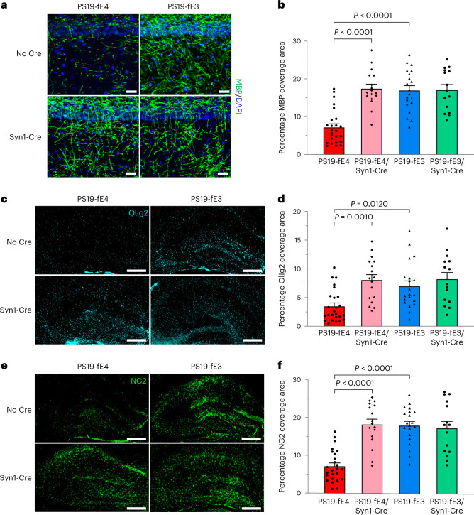
Fig. 3. Myelin deficits and depletion of oligodendrocytes and OPCs are significantly reduced after neuronal APOE4 removal.
a, Representative images of myelin sheath staining with anti-MBP and DAPI in the stratum radiatum of the hippocampus underneath the pyramidal cell layer of CA1 in 10-month-old PS19-fE4 and PS19-fE3 mice with and without Cre (scale bar, 50 µm). DAPI, 4,6-diamidino-2-phenylindole. b, Quantification of the percent MBP coverage area in the hippocampal CA1 subregion of 10-month-old PS19-fE4 and PS19-fE3 mice with and without Cre. c, Representative images of mature oligodendrocytes by immunostaining with anti-Olig2 in the hippocampus of 10-month-old PS19-fE4 and PS19-fE3 mice with and without Cre (scale bar, 500 µm). d, Quantification of the percent Olig2 coverage area in the hippocampus of 10-month-old PS19-fE4 and PS19-fE3 mice with and without Cre. e, Representative images of OPCs by immunostaining with anti-NG2 in the hippocampus of 10-month-old PS19-fE4 and PS19-fE3 mice with and without Cre (scale bar, 500 µm). f, Quantification of the percent NG2 coverage area in the hippocampus of 10-month-old PS19-fE4 and PS19-fE3 mice with and without Cre. For all quantifications in b,d,f, PS19-fE4: No Cre, n = 25; Syn1-Cre, n = 17; and PS19-fE3: No Cre, n = 20; Syn1-Cre, n = 15 mice. All data are represented as mean ± s.e.m., one-way ANOVA with Tukey’s post hoc multiple comparisons test.Source data.

