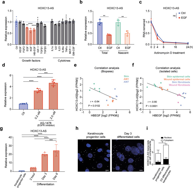
Fig. 3. HOXC13-AS expression is regulated in the opposite direction by keratinocyte growth and differentiation signaling.
a QRT-PCR analysis of HOXC13-AS expression in keratinocytes treated with wound-related cytokines and growth factors for 24 h (n = 4). b QRT-PCR analysis of total and nascent HOXC13-AS in keratinocytes treated with EGF for 8 h (n = 3). c QRT-PCR analysis of HOXC13-AS in keratinocytes treated with EGF and then actinomycin D for 0–24 h (n = 4). d QRT-PCR analysis of HOXC13-AS in keratinocytes treated with AG-1478 for 24 h (n = 4). Expression correlation of HOXC13-AS with HBEGF in the skin and wound tissues (e) and the isolated cells (f) analyzed by RNA-seq. g QRT-PCR analysis of HOXC13-AS in keratinocyte progenitor cells and calcium-induced differentiated keratinocytes (n = 4). Representative photographs (h) and quantification (i) of HOXC13-AS FISH in keratinocyte progenitor cells and differentiated keratinocytes treated with 1.5 mM calcium for 3 days (n = 3). Cell nuclei were co-stained with DAPI. Scale bar = 20 μm. ns not significant, *p < 0.05; **p < 0.01, ***p < 0.001 and ****p < 0.0001 by unpaired two-tailed Student’s t test (a, b, d, g, i), two-way ANOVA (c), or Pearson’s correlation test (e and f). Data are presented as mean ± SD.

