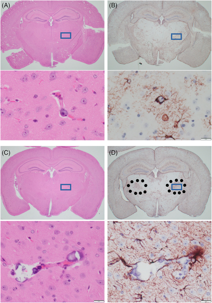
FIGURE 2.

Paraffin sections of FFPE brains of 8‐month (A,B) and 13‐month‐old (C,D) D1113H mice. (A) H&E stain of coronal section of 8‐month‐old D1113H mouse brain. At low power no significant pathology is appreciated. In the inset at high power rare blood vessel in the thalamus shows focal mineralization. (B) In a successive section stained for GFAP symmetric regions of mild gliosis are noted in the thalamus. In the inset at high power these regions show reactive astrocytes surrounding mineralized blood vessels noted in the H&E. (C) H&E stain of coronal section of 13‐month‐old D1113H mouse brain. At low power no significant pathology is appreciated. In the inset at high power blood vessels in the thalamus shows focal vascular mineralization. (D) In a successive section stained for GFAP symmetric regions of gliosis are noted in the thalamus (surrounded by dots). In the inset at high power these regions show reactive astrocytes surrounding mineralized blood vessels as noted in the H&E. Bar in inset = 20 microns
