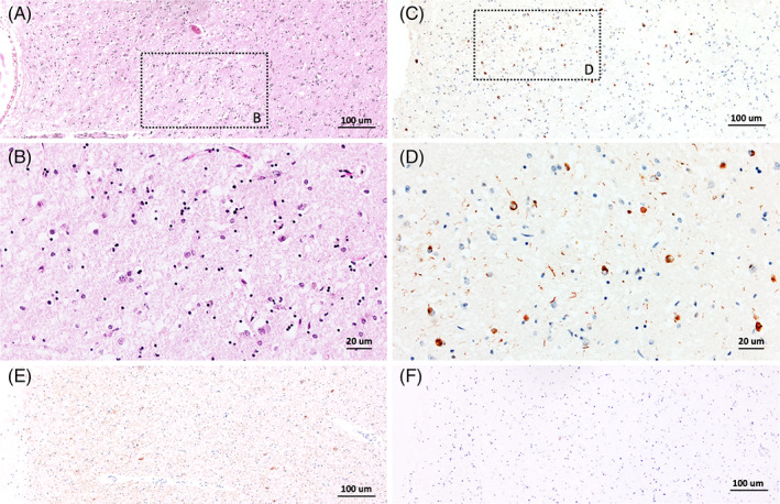
FIGURE 4.

Diagnosis revision in a 90‐year‐old patient with progressive amnestic syndrome. Cortical neuronal loss and gliosis were observed in the hippocampal CA1 subfield (A,B). Phospho‐TDP‐43 cytoplasmic inclusions could be seen in neurons along with phospho‐TDP‐43 immunoreactive dystrophic neurites (C,D). Mild Tau pathology (E) and no amyloid plaques (F) were observed in the same region
