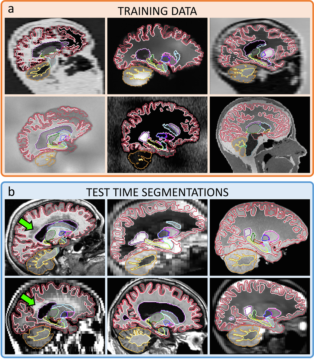
Fig. 1.

(a) Representative samples of the synthetic 3D scans used to train SynthSeg for brain segmentation, and contours of the corresponding ground truth. (b) Test-time segmentations for a variety of contrasts and resolutions, on subjects spanning a wide age range, some presenting large atrophy and white matter lesions (green arrows). All segmentations are obtained with the same network, without retraining or fine-tuning.
