Figure 2.

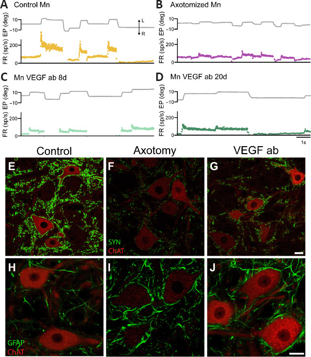
Changes in firing pattern and synaptic inputs in abducens motoneurons treated with anti-VEGF antibody.
(A) Firing rate (FR, in spikes/s) of a control motoneuron (Mn) during spontaneous eye movements (EP, eye position, in degrees). (B) Axotomized abducens motoneurons discharge at remarkably lower frequencies during both fixations and rapid eye movements or saccades. (C, D) Discharge activity of two (intact) motoneurons treated with the neutralizing antibody (ab) against VEGF during 8 or 20 days (C and D, respectively). Note that the blockade of VEGF with the antibody renders motoneurons into an axotomy-like state. The double arrow with L and R in panel A indicates leftward and rightward eye movement, respectively, in A–D. (E–G) Confocal microscopy images of double immunofluorescence in the abducens nucleus combining choline acetyltransferase (ChAT, in red), a marker for motoneurons, with synaptophysin (SYN, in green) to label synaptic boutons. Control motoneurons (E) appear surrounded by numerous synaptophysin-positive boutons. In contrast, after axotomy (F), motoneurons show a massive loss of synaptic boutons as observed by the weak synaptophysin labeling. Uninjured motoneurons treated with the anti-VEGF antibody (VEGF ab) exhibit also scarce synaptophysin-positive boutons (G), lesser than in control (E) and similar to the axotomy situation (F). (H–J) Confocal microscopy images of double immunofluorescence in the abducens nucleus combining (ChAT, in red) with GFAP (in green) as a marker of astrocytes. GFAP immunostaining was very weak in the control situation (H). In contrast, it increased markedly after axotomy (I). In the anti-VEGF antibody situation, although motoneurons are not injured, there was a glial reaction similar to the axotomy condition (J). Scale bars: 20 μm in G for E–G; 20 μm in J for H–J). Reprinted with permission from Calvo et al. (2022). VEGF: Vascular endothelial growth factor.
