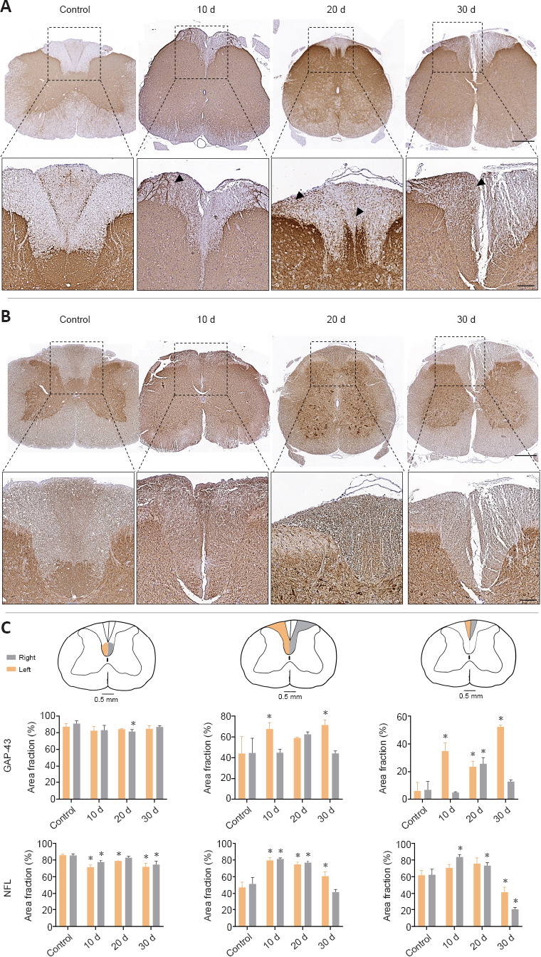
Figure 4.

Immunohistochemical and quantitative evaluation of GAP-43 and NFL expression in the spinal cord after peripheral nerve damage and repair.
GAP-43 (A) and NFL (B) cross-section staining at low and medium magnifications from the central portion of the spinal cord after nerve injury. Arrows indicate GAP-43 positive cells. Quantitative analysis of the right (grey) and left (orange) posterior corticospinal tract, cuneiform fasciculus and gracile fasciculus were included in (C) (data are expressed as the mean ± SD). Scale bars: 500 μm and 150 μm in images with higher magnification. *P < 0.05, vs. control group. Stainings were performed at least three times and in three samples (animals) at each time point as described in the Methods section. GAP-43: Growth-associated protein-43; MCOLL: myelin-collagen histochemical method; NFL: neurofilament.
