
Figure 5.
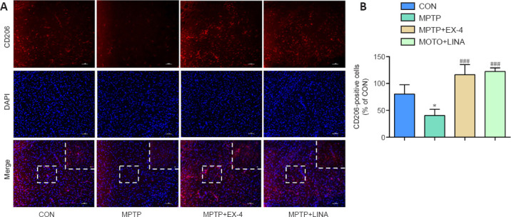
Effects of exendin-4 and linagliptin on microglial polarization in the SN.
(A) Immunofluorescence staining for the M2 phenotype microglia marker CD206 (red, Cy3) in the SN. The intensity of the CD206 signal is weaker in the MPTP group and stronger in the MPTP + EX-4 and MPTP + LINA groups. Scale bar: 50 µm, 20 µm for low and high magnification, respectively. (B) Quantitation of CD206-positive microglia in the SN. Data are shown as mean ± SD (n = 3). *P < 0.05, vs. CON; ###P < 0.001, vs. MPTP (one-way analysis of variance followed by Tukey’s post hoc test). CON: Control; DAPI: 4,6-diamidino-2-phenylindole; EX-4: exendin-4; LINA: linagliptin; MPTP: 1-methyl-4-phenyl-1,2,3,6-tetrahydropyridine; SN: substantia nigra.