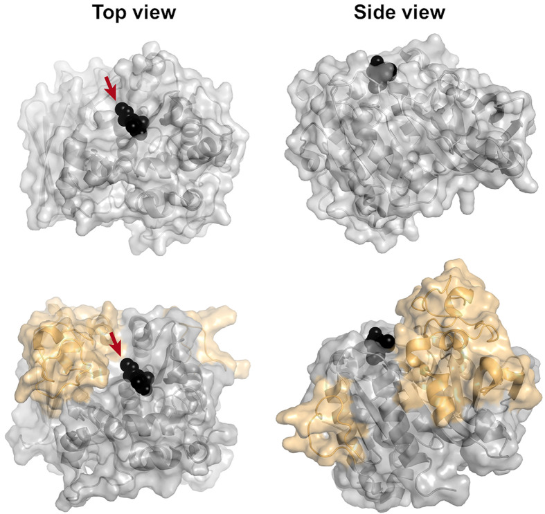
Figure 4. Comparison of fungal and bacterial GE structures.
Top, surface view of the fungal StGE2 (PDB code: 4G4G), and bottom, surface view of OtCE15A (PDB code: 6GRW), illustrating the large inserts relative to the fungal structure, in orange. Me-4-O-MeGlcA bound to StGE2 (and superposed onto the OtCE15A structure) is shown as black spheres. The attachment point for lignin is indicated with arrows.

