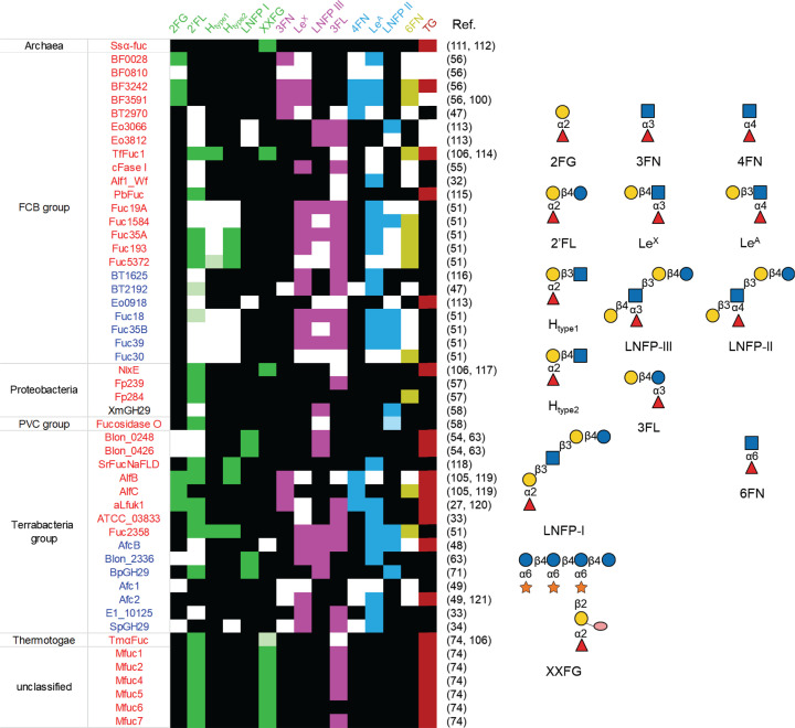
Figure 3. Substrate specificity of microbial GH29 α-l-fucosidases.
The α1,2 substrates are colored in green, α1,3 in pink, α1,4 in sky blue, and α1,6 in olive. Light versions of the above colors indicate trace activity. Black boxes correspond to no enzymatic activity and empty boxes indicate lack of data. GH29A and GH29B α-l-fucosidases are coloured in red and blue, respectively; FCB, Fibrobacteres-Chlorobi-Bacteroidetes super phylum; PVC, Planctomycetes-Verrucomicrobia-Chlamydiae bacterial superphylum; TG, transglycosylation capability. Glycan structures presentation according to Symbol Nomenclature for Glycans (SNFG) [109,110].

