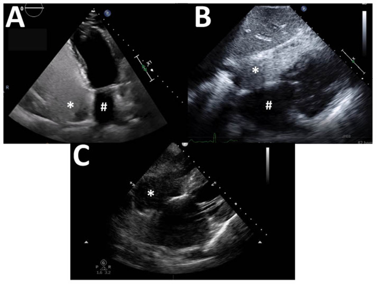
Figure 2.
Common echocardiography views for a transthoracic study. (A) Apical 4-chamber view with left atrium (#) and right atrium (*; densely opacified by agitated saline contrast) in view. (B) Subcostal 4-chamber view with left atrium (#) and right atrium (*; densely opacified by agitated saline contrast) in view. (C) Subcostal 4-chamber view demonstrating inadequate opacification of the right atrium (*); a few scattered bubbles are seen.

