Fig. 5.
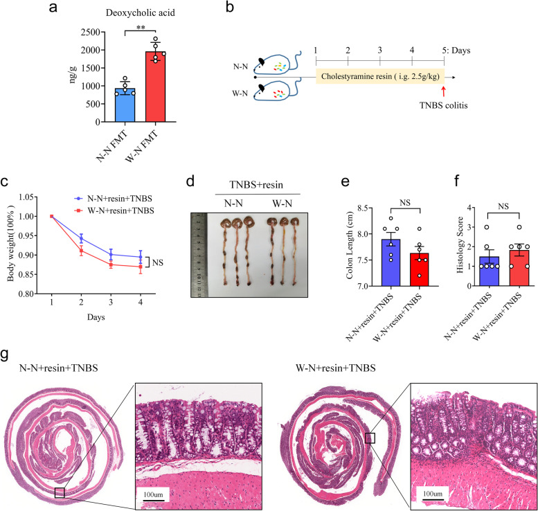
MWD promotes colitis development in their progeny by producing DCA. a As shown in Fig. 2a, WT mice (n = 5) were colonized with gut microbiota from the W-N and N-N groups. Then, the concentration of DCA in the mice’s feces was measured using an ELISA assay kit. b–g Cholestyramine resin (resin) was given to mice in the W-N and N-N groups (n = 6 per group) for 5 days to eliminate intestinal bile acids. Following that, mice were given TNBS to induce colitis. c Body weight changes in mice were evaluated daily after TNBS treatment. d Representative images of TNBS-treated colon in N-N+resin and W-N+resin groups. e The mice were sacrificed on day 4, and the colon length was recorded. f, g Histopathological analysis of colon sections. f Histological scores of colitis were assessed. g Representative images of the H&E-stained colon sections of relevant groups (scale bars 100 μm). a, c, e, and f Data represent means ± SEM; NS, not significant; **P < 0.01; by unpaired Student’s t test. The data shown are representative of three independent experiments

